Your Stroke / Brain Injury Recovery Starts Here


ARNI home-based training and guidance for your rehab is POWERFUL. Accept no substitute.

2026 marks 25 years of ARNI: Click Here to Claim your £50 off Full Set of Stroke Rehab 7 DVDs TODAY!

News

For decades stroke was understood as something that overwhelmingly struck older people, with risk climbing steadily into the seventies and eighties and the under-55s treated as rare, unlucky exceptions, but a run of large studies on both sides of the Atlantic has now overturned that assumption. The most recent, published in Neurology on 23 September 2026 by the American Academy of Neurology, examined records across the Greater Cincinnati and Northern Kentucky region and found stroke in adults aged 20 to 54 had nearly doubled over three decades; 33.9 cases per 100,000 in 1993, rising to 62.2 by 2020, a huge 83% increase.

The same trend has actually already been recorded here in the UK: the Oxford Vascular Study, published in JAMA in 2022, found stroke in the under-55s rose 67% between 2002 and 2018 while falling 15% in those 55 and over, and a much larger record-linkage analysis in PLOS Medicine this year, from the Oxford and British Heart Foundation data teams, found under-55 stroke across England up by around 10% between 2018 and 2024, fastest of all in Asian and Black communities.

In the American data we see that ischaemic strokes accounted for almost all of the rise, while haemorrhagic strokes stayed flat. The study, led by Dr Emily Fisher at the University of Cincinnati, found several risk factors had become more common in the young patients over the years: high blood pressure, diabetes, atrial fibrillation and substance use. It had no healthy comparison group, so it couldn’t establish which of these drove the rise. Differing from the UK, documented substance use climbed from 4.6% to 40.2% across the period, much of it cannabis. Survival improved; deaths within 30 days of a stroke fell from 11.7% in 1993 to 9.4% in 2020.

The Oxford team, led by Dr Linxin Li, found the rise was steepest among people in professional and managerial jobs, which the authors linked to long working hours, stress and low physical activity. The English PLOS Medicine study found undiagnosed high blood pressure and undiagnosed diabetes both rising in the younger patients, and the young were less likely than the old to be on treatment for them. The UCL ULYSSES study, published in the European Stroke Journal in 2025, followed young stroke patients through the North Central London service and found a full half of them (!) still had an unfavourable functional outcome at six months, while 91% reported non-motor problems such as fatigue and disturbed sleep; further papers on their longer-term outcomes have followed this year btw and more to be published next year.

Younger survivors do badly without proper rehab; surviving a stroke at 20 can mean a seventy to eighty YEARS of living with the effects, and those years need very long-term rehab and support, obvs not a few weeks of physio and a discharge letter.

Stroke reaches far younger than any of these studies, too. More than 400 children a year have a stroke in the UK, from babies in the womb through to teenagers, and around 60% of survivors are left with a lasting physical disability. It is one of the leading causes of acquired disability in childhood, but it stays badly under-recognised, and the rehab after hospital discharge is patchy. This is the gap the ARNI Instructors and I are now working to fill. I’m setting up (in collab with one of our amazing stroke survivor donors) a dedicated ARNI Young People’s Stroke Service for the 18-and-under group, with qualified, DBS-checked instructors re-training children in their own homes alongside a parent, and a loan pool of child-friendly rehab equipment. Funding is partly in place.. and fuller details will follow right here on this blog soon! Watch this space 😉
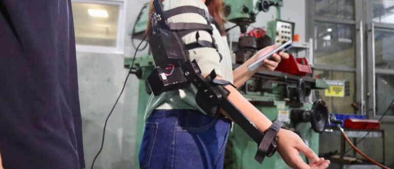
OrthoFlex is a motor-driven elbow exoskeleton built by two University of the Philippines Diliman graduates, Jabez Bote and Ada Koreen Bacay, and named national runner-up in the 2026 James Dyson Award for the Philippines. Its calculated retail cost is about  £130,whereas other home and consumer rehab devices like motorised passive-movement arm trainers, lower-cost rehab gloves, home FES units can start from £350 – and of course clinical exoskeletons start from several thousand pounds to the tens of thousands.

So, as you can see, it targets one joint and one movement; flexion and extension of the elbow, ie bending and straightening the arm. The stroke survivor’s upper arm, forearm and wrist are secured with Velcro cuffs, the device is powered up and paired to a smartphone over Bluetooth, and the user or carer selects flexion or extension through a companion app. A stepper motor drives the elbow through a mechanically limited arc of 0 to 135 degrees, soo it delivers consistent, motor-assisted repetitive elbow practice. OrthoFlex uses a stepper motor driving the joint directly, which is about the cheapest and most easy-to-repair way to move a limb through a set arc.

Much recent academic elbow-exoskeleton work has gone the other way of course, towards sophistication Eg NEUROExos powered elbow exoskeleton, series-elastic cable-driven designs that place compliant actuators along the line of the biceps and triceps, shape-memory-alloy-wire actuators chosen for silent, lightweight operation, and twisted-string actuators that mimic muscle contraction. Those can be back-drivable and compliant, and can run assist-as-needed control that yields to the survivor’s own effort rather than forcing the limb. Each of those refinements adds cost, parts, calibration and fragility. OrthoFlex uses a stepper motor and his/her phone controls the interface work

Two design decisions stand out as rather sensible. The first is the mechanical limit on the range of movement. Rather than trusting software to stop the motor at a safe angle, OrthoFlex caps the arc physically at 0 to 135 degrees; which is good, as the device acts on a stroke-affected joint (which may have spasticity, contracture or altered sensation) So that physical ceiling is a sound, inexpensive safety measure. The second is field-serviceability; the motor housing and cuffs are screwed rather than glued, so worn straps or a failed motor can be replaced part-by-part rather than scrapping the whole unit. Where there is no service centre to post the device back to,home-repairability may be necessary

Set against devices this blog has covered before, the contrast is clear. The VilPower from Vilje Bionics is a whole-arm exoskeleton; ReWalk’s ReStore is a soft exosuit using Bowden-cable actuation to assist the ankle in gait; Kenneth Fong’s powered rehab skateboard at Hong Kong PolyU uses a torque sensor to read the patient’s effort in real time and adjust. All are more capable than OrthoFlex, and all are more expensive and more complex. OrthoFlex competes on access, not capability. Its nearest conceptual cousin is actually the pretty cool open-source work in the field, such as OpenSEA, a 3D-printed series-elastic elbow actuator released openly to lower the barrier to rehabilitation robotics.

This is only a prototype; it of course now needs development and clinical evaluation for safety, usability and whether it produces measurable functional gain rather than simply passive movement alone. The more expensive devices can sense and respond to the patient’s own effort; whether a device this realitevely simple can survivors usefully and actively engaged can only be established by research trial… let’s see what happens.

The NHS is moving its first point of contact from human to machine, and stroke survivors are among the first to find where it fails! The system in the current row about AI is EMMA (Enhanced Medical Management Assistant), a virtual receptionist from QuantumLoopAI, used across many UK GP practices to triage calls, cut queues and handle the ‘8am scramble’ for same-day appointments. A single full-time GP now has well over 2,000 patients, and NHS figures show only about four in ten people are seen on the day they book, so the demand is real. The question is whether an automated voice-recognition layer is the right thing to put between a sick patient and their surgery, and who it shuts out.

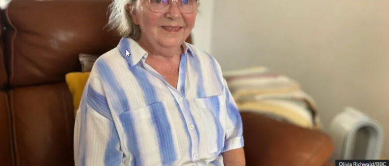
Judith Butterfield, 71, of Armthorpe near Doncaster, had a stroke in January that left her with right-sided weakness and fragmented speech, and spent two months in Doncaster Royal Infirmary. Back home, she tried to book a GP appointment through EMMA five times and couldn’t get it to understand her; upset and frustrated, she felt forced to leave her surgery of long standing and register elsewhere! Apparently, two effects of her stroke defeated the system; it couldn’t make out her fragmented speech and its instruction not to use speakerphone stopped her too, because she holds the handset in her working left hand, which leaves nothing for the keypad her right hand can no longer manage. As she put it: ‘I need to hold the phone in my left hand but I can’t use the keypad now with my right hand, so what am I supposed to do?’

She’s not alone. Healthwatch Rotherham looked into the same system and found it ‘does not respond well to variations in speech such as regional accents or those with speech impediments’, with patients who gave up; one said ‘I could never get it to understand me, I ended up just hanging up’, while others walked to the surgery to be seen in person. Healthwatch England has logged similar problems nationally, mostly among people with speech or learning difficulties. The people it fails are the very same people that a stroke, or ageing, or neurological disability most affects….

There are two separate problems. One is accent and dialect coverage, which a vendor can improve with better training data. The other is disability, and the reasonable adjustments the law requires for it. Dysarthria, aphasia and hemiparesis are absolutely conditions the NHS exists to accommodate so  the anodyne ‘Ask for a human at any time’ doesn’t solve anything at all. The NHS Accessible Information Standard has required practices to meet disabled patients’ communication needs since 2016; and yet the NHS App is set to use AI to triage patients in a roughly £10 billion overhaul, adopted practice by practice rather than nationally. What do you think? Will this create a better service or just more a whole lot more frustration?

If your arm and hand have been left weak by a stroke, you’ll know it’s often the last thing to recover, and standard rehabilitation has surprisingly little to offer once you plateau. So this pilot trial from the University of Pittsburgh, published in Nature Medicine on 4 June 2026, caught my eye. Seven people with chronic, profound arm weakness got a measurable amount of movement back… using an implant borrowed from a completely different field of medicine.

As you know so well, a stroke damages the brain, not the spinal cord, and that turns out to be the whole basis of the idea. When you go to move your arm, the command still leaves the brain and travels down through the spinal cord to the neurons that fire the muscles; after a stroke those descending signals are badly weakened, but the spinal circuits below are still intact. They’re simply not getting a strong enough command any more. As lead researcher Marco Capogrosso puts it: ‘we’re not substituting anything. We’re just trying to facilitate whatever is left after stroke and help it as much as we can.’

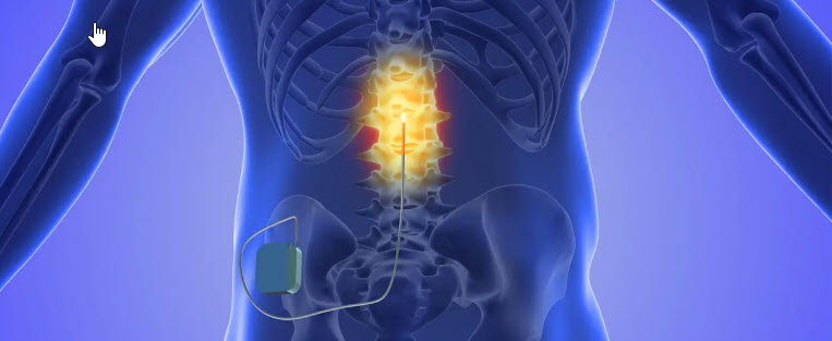
The technique is called cervical epidural spinal cord stimulation. Two thin electrode leads are placed just outside the spinal cord at the neck, over the roots that serve the arm and hand (C3 to T1). The stimulation doesn’t drive the muscles directly; instead it targets the sensory nerve fibres entering the spinal cord (the dorsal-root afferents), and through them raises the excitability of the motor neurons, so the weakened signal still arriving from the brain is now enough to produce movement. The same class of device has been used for decades to treat chronic pain, and is already FDA-approved for that, but this is the first time anyone’s used it to restore arm function after a stroke.

The results, for a small early trial, were well worth having. All seven participants improved, whatever their starting level: an average 32% increase in arm strength, better overall arm mobility, and a reduction in the spasticity (the abnormal muscle stiffness) that so often follows stroke. And all of this came from fewer than nine hours of movement training across four weeks, with no serious side effects and no pain. One participant’s goal was simple: ‘my goal in the beginning was to be able to hug my girlfriend and daughter with both arms. I was able to do that.’ Professor George Wittenberg, a co-author, didn’t hold back: ‘it is like a quantum change in the way that stroke rehabilitation is being handled.’

Two things stand out about it. First, the effect is immediate; switch the stimulation on and the movement improves that moment, which, as Capogrosso notes, is itself a real motivator, because the survivor sees and feels the difference straight away and engages more. Second, it isn’t purely switch-on-switch-off. The stimulation gives an assistive effect while it’s on, but the research also points to a therapeutic effect that builds over time, with some improvement remaining even when it’s off. Btw, the one honest caveat from this feasibility study is that holding onto the gains largely depended on continued stimulation; function declined when the stimulation stopped, which is one of the main questions the next trial is set up to answer.

There’s real reason to take this seriously beyond the small numbers. Around 400,000 people in the US develop chronic arm and hand weakness after stroke every year, and, as Wittenberg says, we’re increasingly comfortable with implanted devices anyway: ‘people already get pacemakers to keep their heart going, so why not have a spinal cord stimulator to keep their movement going?’ The team is now recruiting for a longer trial, testing extended stimulation both on its own and alongside physical therapy, and Capogrosso reckons that, if no major obstacles come up, the technology could be approved for stroke within three to five years. That’s US-based and still some way from your local hospital here of course… but as timelines for this kind of neurotech go, three to five years is not long at all.

At ARNI, the ARNI Instructors and I have always worked on the principle that the arm comes back through intensive, repeated, meaningful practice… and nothing here changes that. What something like this offers is a way to make that practice possible for people whose signal to the muscle is currently just too weak to act on. Interesting stuff, and we’ll be watching where the next trial takes it.

After a stroke, a common cause of a stiff, hard-to-control upper limb is something called abnormal co-activation, or coupling; muscles that should fire separately instead fire together, so when you try to do one movement you might get an unwanted second one bundled in with it. The brain’s movement signals have, in effect, become ‘tangled’. A team at Northwestern University, led by Professor Marc Slutzky, has built a retro, 90s-style video game to tackle this effect, and published the results in Neurorehabilitation and Neural Repair in June 2026.

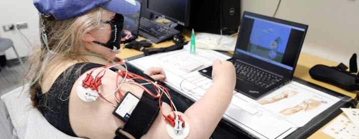
Slutzky’s system, called MINT (myoelectric interface for neurorehabilitation) works like this. You wear a small device on your affected arm that reads the electrical activity of your muscles (the electromyogram, or EMG). Two coupled muscles are each mapped to a direction on screen; say, your biceps moves the cursor right and your deltoid moves it up. If those two muscles are abnormally coupled, they fire together, so the cursor only travels diagonally, up and right at once, like turning both dials on an Etch-a-Sketch simultaneously. The game then places targets further and further off that diagonal, so the only way to reach them is to activate one muscle without the other. To score, you have to decouple. The more the muscles separate, the higher you score, and the separating is the therapy.

The results were strong, though the headline number needs a caveat. Participants were chronic and severely impaired; on average 6.4 years past their stroke, some as much as 12 years out, able to lift the arm a little but not straighten it. After six weeks, the best-performing group improved arm function by 7.8 times as much as a sham control group; to be precise – the impovement group being the group which was given an extra week of training on the most impairing muscles. Across the experimental groups as a whole, the improvement was around 4.5 times the sham group, which the researchers still rate as clinically meaningful. Two further points stand out; the survivors kept improving even after the therapy stopped, and there was a direct correlation between how much the co-activation reduced and how much movement improved, which points to the decoupling as the active ingredient rather than the exercise or the fun of the game.

The access angle is what makes it practically interesting.. because one can play at home, the training volume goes up sharply. Participants managed more than 300 repetitions a day, against perhaps 30 reps, three days a week, in typical clinic-based therapy. ‘Dosage’ sufficiency is acknowledged by experts in upper limb rehab such as Professor Nick Ward at Queen Square, UCL, to be one one of the persistent problems in stroke rehab – he says that most survivors end up getting a ‘homeopathic dose of training’ during their clinical rehab time. A cheap wearable plus a laptop would most probably a survivor rack up reps really easily. It also treats the impairment directly, rather than teaching the survivor to compensate (leaning the whole body forward to reach, say, instead of extending the arm); useful as compensation is, it actually doesn’t improve the arm itself. Slutzky’s team is apparently making the wearable fully wireless, upgrading the games to be more engaging, and is planning to test the same principle on survivors’ lower limbs.

A large stroke often leaves a permanent hole in the brain. Once the dead tissue is cleared away you’re left with a cavity, and up to now that’s been seen as permanent damage that can’t be undone or resolved, with rehab as the only possible solution. But a team at Duke University, led by Professor Tatiana Segura, has been trying something different to past attempts to ‘fill the gap’; injecting a smart hydrogel into that cavity to turn it into a place where the brain can start repairing itself. Basically, the gel is injected as a liquid, then sets into a soft, sponge-like scaffold full of tiny interconnected pores. The pores give the body’s own cells somewhere to move into and start rebuilding, and the gel also softens the dense scar that normally seals off the damaged area. So instead of just filling the hole, it opens it up as a sort of building site for repair.

The scaffold also carries tiny biological ‘signal packages’ (taken from astrocytes, the brain’s star-shaped support cells) to attract the immune cells that help with healing. Injecting those signals on their own did almost nothing; they only worked when anchored into the gel, where arriving cells could find them. The results are in Cell Biomaterials (July 2026) – and it shows that the effects in mice were strong. New blood vessels grew through the cavity, fresh nerve fibres (the wiring that lets brain cells connect) appeared, and, most importantly, movement improved. Mice given the gel did much better on a walking-coordination test, and after eight weeks they were performing on a par with healthy animals. It also worked when given more than 24 hours after the stroke; well outside the tight window that clot treatments need.

The idea of filling the stroke cavity isn’t new in and of itself btw; researchers have been trying to put biomaterials into that empty space for well over a decade now. Segura’s own earlier gels (developed first at UCLA) showed back in 2017 and 2018 that an injectable hydrogel could reduce scarring and draw new blood vessels and young nerve cells into the cavity in mice. Others have tried loading similar gels with growth factors, with stem cells, or with drugs, using the scaffold as a way to hold treatment in place at the injury rather than let it wash away through the bloodstream. So the cavity as a space to build in is a fairly established line of research.. what has varied to date is the various proposals re what you put into the scaffold, and how you get the brain to respond to it.

What’s harder, and what most of these attempts have struggled with, is turning that early promise into lasting, useful movement. Plenty of methods can grow a few new blood vessels or nudge some cells to migrate; far fewer produce the coordinated rebuilding (vessels, nerve fibres and the right immune activity all together) that actually shows up as better function. That’s the part this latest work is concentrating on, and it’s why the immune-system angle is drawing interest. So, the earlier gels mostly acted as passive scaffolding… whereas this one apparantly tries to organise the repair response around it. Whether that holds up beyond mice is the open question, but it’s a reason the field keeps coming back to the cavity idea despite the slow going. This is early animal research but the idea is a hopeful one for those of us dealing with long-term damage; not restoring blood flow (too late for that), but I think preparing the old, settled injury so the body can rebuild, and giving stroke rehab trainers more to work with is a great target to work for.

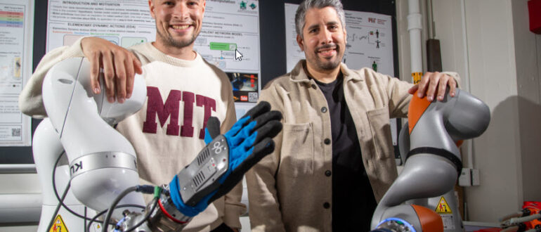
Most ‘rehabotics’ (rehab-robotics) so far has been good at one thing and poor at another. It can move a limb along a set path well enough, but it really struggles with the part a human therapist would do instinctively; sensing how much resistance to give, when to yield, when to push and adjusting all of that moment to moment by feel, etc. A group at MIT, working with the Technical University of Munich, has built a system aimed at exactly that gap. The team (Johannes Lachner, now at Purdue, and Noah Geiger, with Neville Hogan’s Newman Laboratory at MIT) set a clear design principle; extend therapists, not replace them, and let a therapist train their own robot. Their paper, “Diffusion-Based Impedance Learning for Contact-Rich Manipulation Tasks,” is in IEEE Transactions on Robotics.

The method fuses two established ideas. The first is impedance control, a long-standing robotics approach (Hogan’s own field) that governs how a robot responds to physical contact by setting its stiffness and damping; how rigidly or softly it reacts to being pushed. It’s stable and safe, but an engineer has to ‘hand-tune’ stiffness and damping values for each task, which doesn’t scale to real, varied rehab. The second is a generative diffusion model, the same class of AI used to generate images, but here it’s repurposed to generate behaviour rather than pixels.

What the diffusion model produces works like this: from the forces measured at the robot’s contact point (the external ‘wrenches’), a transformer-based diffusion model reconstructs what the researchers call a simulated Zero-Force Trajectory; the path the limb would follow if it were moving freely, with no resistance. An energy-based estimator then compares that intended path with what’s actually happening under contact, and adapts the robot’s stiffness and damping in real time, tightening along the direction of the task and softening along the axes that are irrelevant to it. So instead of a fixed, pre-tuned response, the assistance is continuously being ‘reshaped’ by the forces the patient is producing.

The performance figures show how well this works. Trained on only tens of thousands of samples, gathered by teleoperating the robot through both rehabilitation movements and an obstacle ‘parkour’ using an Apple Vision Pro headset, the model reached sub-millimetre positional and sub-degree rotational accuracy, ran in real-time torque control on a standard KUKA LBR iiwa arm, and generalised to tasks it had never seen; a 30/30 success rate inserting cylindrical, square and star-shaped pegs it wasn’t even trained on. In other words, it had learned something about physical interaction, not just memorised a set of movements – which I think is quite amazing.

In an ongoing study with Professor Cristina Piazza’s group at TUM and the Pfennigparade Rehabilitation Centre in Munich, physios and OTs are treating patients while wearing force-sensing gloves and being filmed. That data is being used to build models that capture the physical style of an individual therapist, with the eventual aim of therapist-specific models, evaluated against the same patients who previously had manual therapy. So the robot learns from expert human handling rather than from an abstract idealisation of it,  then delivers high-volume, force-adaptive practice to stroke survivors under supervision… this seems to me to be to be very useful – and on the right track to make robotic additions actually helpful addition, rather than promising so much and actually not really delivering anything much more than a therapist can do very well a without vastly expensive robot…

Peter Coghlan’s recovery story is one of the more remarkable you’ll come across. In 2011, aged just 33, this former soldier had a massive brain stem stroke and woke from a coma with locked-in syndrome; fully conscious and aware, but unable to move or speak. He could blink, and that was all. Imagine lying there, hearing people around your bed discuss a bleak future, with no way but blinking to tell them you’re still in there….

Locked-in syndrome is caused by damage to the pons, the part of the brain stem that carries nerve traffic between brain and body. Almost every voluntary muscle is paralysed, except, usually, the ones that move the eyes; so the person is awake and thinking clearly but unable to move or speak. Around 90% of people with it die within four months, and there’s still no cure, only assistive technology to help with communication.

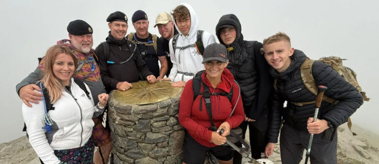
Peter actually learned to communicate letter by letter on an alphabet board, using only his eyes. His first four words, blinked out to neurologist Professor David Blacker, were: ‘can you fix me?’ Then came the work; hours every day, trying to move a finger, trying to swallow, relearning the basics most of us never think about. At times, he says, it was like a horror film. But he kept at it. Six months and one day after his stroke, Peter walked (when no-one with his condition had been known to walk independently out of a hospital ward). Since then he’s kept setting himself challenges; 7km of Perth’s City to Surf, 15km for a charity walk, and last year he hiked Snowdon, the highest mountain in Wales, raising over £500 for fallen soldiers and quite possibly becoming the first locked-in syndrome quadriplegic stroke survivor to do it. He now volunteers on the same stroke ward where he was once a patient, and he’s written a book about it all, In the Blink of an Eye. Amazing stuff 😉

A word of caution alongside the inspiration, as ever. Peter’s degree of recovery is really rare; most people who survive locked-in syndrome don’t regain anything like this, and it depends heavily on the site and extent of the brain stem damage and much else outside anyone’s control. So it’s not a template; but it does show what can happen when someone refuses the ceiling placed on them and keeps chipping away long after most would have stopped, and that’s exactly the principle the ARNI Instructors and I work on every day; there’s often more in the tank than the first prognosis suggests. Peter did it with an alphabet board and a refusal to give in – great job Peter.

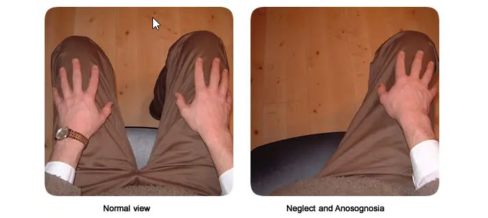
If you’ve had a stroke on the right side of the brain, you may well have come across a problem called spatial neglect, which is where the brain stops attending to everything on your left. It isn’t actually blindness; your eyes work perfectly well, but the brain no longer registers one side of the world. Survivors tend to miss food on the left of the plate, walk into door frames, read only the right half of a page – or don’t see a car coming from the left at a crossing. Neglect is among the most common cognitive effects of stroke, occurring in roughly 38% of survivors, and it’s also one of the strongest signs of a slower, harder recovery, so finding it early can change what comes next.

The trouble is that the usual bedside tests miss a good deal of it. Someone can complete a pen-and-paper cancellation task, or copy a drawing well enough, and still be neglecting their left side out in the daily life where the problem really bites. Conventional screening depends on structured verbal instructions and set tasks, which don’t always reflect how a person behaves when simply looking around a room. And, for survivors whose speech has been affected by the stroke, language-based tests become even harder still to administer fairly.

So a team at the University of Lucerne and the Lucerne Cantonal Hospital, led by Prof Thomas Nyffeler and Dr Brigitte Kaufmann, has spent several years developing a different approach based on eye-tracking, and their latest study (published in Neuropsychological Rehabilitation in 2026) takes it a significant step closer to everyday clinical use. The method is called free visual exploration. You simply look at photographs of ordinary everyday scenes, and an eye-tracker logs where your gaze goes. Someone with neglect explores only the right side of each image and leaves the left almost untouched, and when a clinician maps that gaze as a heat map, the imbalance shows up plainly. As you might imagine, this is far closer to how neglect actually behaves in real life than a tick-box test at the bedside!

What the new study adds is the missing piece that has held eye-tracking back from routine use: proper reference data. The team measured healthy people aged 20 to 89 to establish what normal looks like at every age, so a clinician now has age-appropriate benchmarks to compare a patient against. The overall direction of gaze stayed stable right across adulthood, which means the result holds up whatever a person’s age. When they tested the benchmark from their earlier work, it correctly identified over 95% of healthy participants, so a clinician can read an unusual result with real confidence as a genuine sign of neglect rather than ordinary variation in how people look at a scene. And their earlier research had already shown that clinicians pick up neglect more reliably using free-exploration eye-tracking than with the standard paper-pencil tests.

There are practical reasons this suits stroke survivors particularly well; it’s quick and needs almost no spoken instruction, so it works whatever a person’s first language, and even where speech has been affected by the stroke. And because you can repeat it easily and it gives consistent results over time, a therapist can use it not just to diagnose neglect once, but to track whether it’s improving as rehabilitation goes on. To help other clinics take it up, the team has made their normative data and analysis scripts openly available, so hospitals and neurorehabilitation centres can apply the same benchmarks straight away. It’s still moving from research into routine practice rather than sitting in every stroke unit today, but that transition looks a good deal closer than itdid. So a fast, language-free way for clinicians to pin it down early, and then follow it through recovery, is a welcome direction… and one we’ll be watching closely as it reaches more UK clinics.

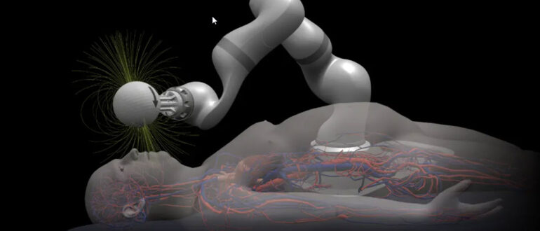
How about this; imagine a soft, hair-thin thread being steered through the twisting blood vessels of the brain by a magnet held outside the skull, clearing the clot that caused a stroke. Science fiction, right?! But amazingly, it’s being built right now, by a team at MIT and a spinout called Magnendo led by Dr Yoonho Kim. And it could change how the most severe strokes are treated, and crucially, where they can be treated.

As you may know, when a large clot blocks one of the major arteries supplying the brain, the treatment is a thrombectomy: a surgeon threads a thin guidewire up through the blood vessels, from an artery in the groin or wrist all the way to the brain, then passes a catheter over it to pull the clot out. It’s one of the most effective treatments in all of stroke medicine. The hard part is the journey. The brain’s vessels are narrow, winding and fiendishly awkward to navigate, and steering a passive wire around those bends by hand takes real skill, real time, and carries a genuine risk of damaging a vessel wall along the way.

Magnendo’s answer grew out of years of research at MIT into what the team call ferromagnetic soft continuum robots. The guidewire has tiny magnetic particles built into a soft polymer tip, so instead of being pushed and twisted by hand, it’s steered from outside the body by a magnet mounted on a robotic arm. Tilt the magnet, and the tip follows the magnetic field around even the sharpest corner. In their earlier work, published in Science Robotics, the team showed the wire threading through a life-sized silicone model of the brain’s vessels, making one acute-angled turn after another to reach a series of aneurysms.. the wire is actually as thin and flexible as a conventional neurovascular guidewire, but where a standard wire is passive, this one actively steers itself toward the target.

The approach is posited to be able shorten the navigation stage of the procedure by 10 minutes or more in a typical case, and up to 30 minutes in a complex one, which is critical stuff when brain tissue is dying by the minute. In preclinical testing it reached hard-to-reach spots faster and more consistently, and, tellingly, it narrowed the gap between more and less experienced operators. The team has now been backed to develop the system further, with the longer-term aim of it navigating largely on its own, guided by live imaging and AI, so that a surgeon supervises rather than manually threads every millimetre.

So what could this mean for us as survivors and families? I think first about speed. Every minute a major artery stays blocked, more of the brain is lost, so anything that gets the clot out sooner means less damage and, very often, a better recovery to build on afterwards. But the bigger prize is access. Thrombectomy is brilliant, yet right now it’s only offered at a small number of specialist centres, because it needs a highly trained expert on hand, and many people who could benefit live too far from one to reach it in time. A robot that can do the difficult navigation, and could one day be operated remotely, might bring this treatment within reach of far more hospitals, so that where you happen to live weighs less heavily on whether you get it.

This is really early-stage work; larger trials and a long road through regulation lie ahead before any of it becomes routine. But the direction is a hopeful one: faster, safer clot removal, potentially available in many more places, for many more people. And obviously, the less brain tissue lost in those first crucial hours, the more there is to work with in rehabilitation afterwards… sounds good to us at ARNI Stroke Rehab… what’s your view?


We are on Facebook

ARNI