Your Stroke / Brain Injury Recovery Starts Here


ARNI home-based training and guidance for your rehab is POWERFUL. Accept no substitute.

2026 marks 25 years of ARNI: Click Here to Claim your £50 off Full Set of Stroke Rehab 7 DVDs TODAY!

News

Measuring stroke recovery properly is harder than it sounds. Most single outcome measures capture one dimension of what stroke does to a person and miss everything else… which is why Dr Tom Balchin at ARNI Stroke Rehab UK has built a set of four complementary assessments into every ARNI specialist’s training and into the Training Logbooks that survivors and instructors use together, repeated every 12 weeks so that genuine progress – and any regression – can be tracked with rigour over time.

The Stroke Impact Scale (SIS) is introduced from day one of the ARNI Functional Rehabilitation after Stroke Accreditation; it covers strength, memory, emotion, communication, activities of daily living, mobility and quality of life, giving both trainer and survivor a rounded picture of how stroke is affecting daily living across every domain. Alongside it, also from the outset, comes the Chedoke Arm and Hand Activity Inventory (CAHAI); it assesses the functional use of the affected arm and hand across nine real-life bilateral tasks — opening a jar, doing up buttons, pouring a glass of water and more – producing a score out of 63 that reflects what the upper limb can actually do in practice rather than just what it looks like in a clinical setting. Together the SIS and CAHAI form a strong and complementary pairing; the SIS capturing the broad whole-life picture and the CAHAI drilling down into the upper limb detail that sits at the heart of so much of ARNI’s rehabilitation work.

Once students are established in the course, the Rivermead Mobility Scale (RMS) is introduced; it measures mobility across 15 tasks from turning over in bed through to running, making it an ideal tool for tracking lower limb and whole body functional progress over time. And most recently Dr Tom has begun introducing instructors to the Fatigue Severity Scale (FSS), developed by Krupp et al… which was advised to him by his kind contact Dr Anna Kuppuswamy (formerly of Queen Square, UCL and now at Leeds), because post-stroke fatigue is one of the most debilitating and underassessed sequelae of stroke, recognised by survivors themselves as something that can affect them every day.

What seems to make this quad quite effective is that together they cover the full landscape of stroke recovery without much overlap – upper limb function, mobility, whole-life impact and fatigue – and no single measure could do that alone. They are the evidence base that shows survivors, families and commissioners what ARNI training is actually achieving, session by session and month by month. Every ARNI instructor is trained by Dr Tom and his team to use them with care and consistency.

Stroke prevention research tends to focus on the dramatic; new drugs, surgical interventions, advanced imaging – and it’s easy to overlook the quieter evidence building around diet. A study published in March 2026 in the journal Nutrients by Wakayama, Araki, Nakamura and Ikeda shifts attention toward one of the most ordinary dietary habits imaginable: drinking milk. The projected results are striking enough to deserve serious attention from anyone interested in the population level reduction of stroke burden.

The research used a Markov model to simulate how a population moves between health states over time when a risk factor changes; each annual cycle in the model tracked individuals moving between no stroke, stroke, stroke-related death and death from other causes. It modelled the effects of increasing milk consumption to the nationally recommended 180 grams per day among Japanese adults aged 30 to 79 over ten years, examining stroke incidence, stroke-related deaths and national healthcare expenditure. Two scenarios were tested – an immediate increase and a gradual one building at a constant annual rate – and both produced meaningful projected benefits, though the immediate change performed better across most outcomes. Meeting the recommended dairy intake was projected to reduce stroke incidence and stroke-related mortality by approximately 7% overall, with some subgroups seeing reductions of up to 10.6%… and stroke-related national healthcare expenditure was projected to fall by around 5.1%, with subgroup savings reaching as high as 8.5%. The greatest absolute benefits appeared in older adults aged 70 to 79, while younger groups showed the largest proportional improvements. And crucially, the dietary change being modelled is simply meeting an existing national recommendation that most Japanese adults currently fall short of.

The biological plausibility is reasonably well established even if the precise mechanisms remain incompletely understood. Milk provides calcium, magnesium and potassium, all of which have been associated with blood pressure regulation and cardiovascular protection; and hypertension is the single most important modifiable risk factor for stroke, so any dietary pattern that plausibly contributes to lower blood pressure deserves attention in this context. The association between dairy intake and reduced stroke risk has been examined across multiple study designs and populations, and the direction of the evidence is consistent… a large dose-response meta-analysis drawing on 18 prospective cohort studies covering over 762,000 individuals and nearly 30,000 stroke events found that each additional 200 grams of daily milk intake was associated with a 7% lower risk of stroke, with the association particularly strong in East Asian populations. A separate global analysis published in Nature Communications in 2025, drawing on both the China Kadoorie Biobank and the UK Biobank, found that total dairy consumption was associated with a 6% reduced risk of stroke across the combined dataset.

It’s important to be clear about what this Nutrients study is and isn’t. It is a simulation rather than a randomised controlled trial demonstrating that increasing milk intake causes a reduction in stroke events… the model cannot account for every confounding variable, it doesn’t estimate the costs of implementing population level dietary change, and it doesn’t disaggregate by milk type or stroke subtype in ways that would allow more precise policy recommendations. Future studies could usefully incorporate long-term caregiving costs, examine differences between whole and lower fat milk, and look more carefully at hormonal status and age related differences to generate more precise estimates.

Japan’s dietary context also differs meaningfully from the UK’s, and the specific projections shouldn’t be transplanted directly into a UK policy context without replication in relevant cohorts. But the broader message is clear enough to take seriously now. Diet is a modifiable stroke risk factor, the evidence for dairy intake as part of a holistic prevention approach is accumulating steadily, and translating findings like these into UK public health guidance requires review by bodies such as NICE and the British Dietetic Association — a process measured in years rather than months. So… the time to be paying attention is now, not later!

The University of Manchester has spent the better part of two decades building the case for anakinra (IL-1Ra) as a treatment for ischaemic stroke – and the latest research from the group, published in the American Heart Association’s journal Stroke and funded by the Medical Research Council, finally explains why a drug with such strong early promise failed to deliver in clinical trials. The answer has less to do with the drug itself than with when it was given.

Stroke is the second leading cause of death and disability worldwide, and despite decades of research tPA remains the only licensed thrombolytic treatment for ischaemic stroke in the UK, given within a strict 4.5 hour window and received by only around one in ten patients. The biological case for anakinra is well established; interleukin-1 drives inflammatory responses in the brain after stroke, attracting white blood cells that kill nerve cells and worsen injury rather than helping, and blocking IL-1 limits that secondary damage. Early animal studies were striking, with rats given IL-1Ra showing roughly half the brain damage of controls… but the phase II SCIL-STROKE clinical trial, based at the Northern Care Alliance NHS Foundation Trust, failed to show overall improvement in patient recovery.

Lead author Dr Ioana-Emilia Mosneag and colleagues at Manchester re-examined the trial data and found that nearly three-quarters of SCIL-STROKE patients had received tPA before IL-1Ra, and those patients had significantly lower levels of IL-1Ra in their blood- the drug was being broken down. Laboratory work confirmed that IL-1Ra is cleaved by plasmin, the enzyme produced during tPA treatment, meaning the anti-inflammatory drug was being degraded before it could work… and mouse model experiments made the scale of the problem clear. When given simultaneously with tPA, brain damage reduced by only 15% compared to 68% with tPA alone. But when IL-1Ra was given after tPA, no harmful interaction occurred and the protective effects of tPA were fully preserved.

As Professor Stuart Allan noted, ‘timing is very likely to be a critical factor in the efficacy of IL-1Ra, which will be beneficial if given after tPA rather than alongside it.’ Professor Craig Smith added that future studies will need to investigate the timing and effectiveness of IL-1Ra after tPA, and whether similar interactions occur with tenecteplase, a newer thrombolytic increasingly used in UK stroke centres that may be less likely to degrade IL-1Ra due to its greater specificity. Routine clinical implementation remains some years away pending larger human trials and NICE review; but the failure of SCIL-STROKE was not a dead end – it was a timing problem, and that is a very different thing.

The clinical management of post stroke cognitive impairment has historically been complicated by the lack of reliable predictive instruments capable of addressing the heterogeneous nature of neurological recovery. To resolve this significant gap in care researchers at the Nuffield Department of Clinical Neurosciences at the University of Oxford have developed a novel Stroke Cognition Calculator designed to provide earlier and more informed estimates of long term thinking problems. Funded by the National Institute for Health and Care Research and published in the Lancet Health Longevity this study addresses the ‘invisible’ challenges such as memory deficits and impaired decision making that frequently hinder a survivor’s return to independence. The tool utilises data already captured during routine hospital admissions including patient age and stroke severity alongside results from the Oxford Cognitive Screen which is a bedside assessment already integrated across the National Health Service. By synthesising these variables the model offers a personalised outlook that helps families and clinicians move beyond the uncertainty that often characterises the transition from hospital to home.

The empirical strength of the Stroke Cognition Calculator lies in its superior accuracy when compared to existing prognostic models. During initial validation with 430 participants in Oxford the tool achieved a 76 per cent accuracy rate in predicting cognitive status at the six month milestone. This performance significantly outperforms previous tools which typically recorded accuracy rates between 53 and 66 per cent. A critical technical distinction is that while prior instruments focused almost exclusively on predicting cognitive decline the new calculator accounts for the dynamic nature of recovery where cognition may improve or remain stable. Further multi centre testing involving 264 participants across 37 hospitals in England demonstrated that the accuracy remained robust at 74 per cent even when applied in varied healthcare settings. This high level of generalisability suggests that the tool is ready for broader clinical implementation as a means of ensuring equitable access to tailored support services.

First author & colleague of ARNI Stroke Recovery UK, Andrea Kusec, has noted that the high potential for everyday clinical use stems from the fact that the calculator relies on predictors already present in medical records making it both affordable and practical to deploy. Professor Nele Demeyere highlights that while cognitive difficulties are common and varied the ability to predict those who will continue to struggle is a major step toward more personalised post stroke care. By identifying individuals at higher risk of persistent impairment clinicians can strategically allocate resources and provide families with realistic expectations of the recovery journey. ARNI Stroke Rehab UK says that providing survivors with an accurate understanding of their cognitive trajectory is pretty essential for maintaining the mental resilience required for physical retraining… and that this innovative prediction tool could possibly help with the integration of cognitive support to  the intensive task specific training programmes that are vital for regaining functional independence in the community.

A pioneering study published in the journal Nature has detailed the development of a magnetic hydrogel designed to be injected into the heart to function as a biocompatible plug. This is a bioengineering solution that targets the left atrial appendage (a common site for thrombus formation in patients with AF). This innovative substance is formulated to remain fluid during the injection phase but undergoes a rapid phase transition to a solid state once positioned within the target cardiac architecture. By occluding the left atrial appendage, the hydrogel effectively prevents the migration of blood clots into the systemic circulation, thereby reducing the risk of ischaemic events. Research conducted on animal models, specifically rats and a porcine subject, has demonstrated that the gel adheres securely to the heart wall without causing significant inflammation or structural damage.

The technical complexity of this magnetic gel allows for precise positioning using external magnetic fields, ensuring that the occlusion is complete and stable over time. Traditional methods for closing the left atrial appendage often involve invasive surgical procedures or the permanent implantation of metallic devices which can carry risks of erosion or incomplete sealing. This hydrogel alternative offers a minimally invasive pathway that could potentially be performed under local anaesthetic, significantly expanding the eligibility criteria for elderly or frail patients. Furthermore, the material is engineered to integrate with the surrounding cardiac tissue, promoting a natural healing response that further stabilises the blockage.

As the global burden of stroke continues to rise, such biotechnological advancements provide a critical second line of defence for those who cannot tolerate long term anticoagulant therapy. The integration of these advanced materials into human clinical trials will definitely require rigorous assessment of long term stability and potential degradation products within the bloodstream. Initial results suggest that the magnetic properties don;t interfere with standard imaging techniques like magnetic resonance imaging.. which is a vital consideration for future patient monitoring.

Hyun, J., Kim, J., Wang, S. et al. (2026). Magnetogel for minimally invasive occlusion of the left atrial appendage. Nature, 639, 94–101.

The clinical and legal implications of misdiagnosed cerebrovascular accidents in the United Kingdom have been brought into sharp focus by a significant settlement involving the Cambridge University Hospitals NHS Foundation Trust. In a case that exemplifies the catastrophic consequences of diagnostic inertia, a stroke survivor from Bishop’s Stortford was awarded one million pounds in compensation following a series of systemic failures at Addenbrooke’s Hospital in 2016. The patient, identified as Lisa, presented with acute left-side paralysis… a classic red-flag symptom of an ischaemic event – yet was not referred to a stroke specialist. Following an eight-hour wait and an inconclusive CT scan, clinical staff erroneously attributed her neurological deficits to stress rather than vascular pathology. This failure to implement the standard stroke pathway led to her premature discharge in a wheelchair, despite her inability to ambulate independently.

The subsequent clinical trajectory highlights the ‘window of opportunity’ lost when initial assessments are flawed. While at home, Lisa suffered a second, more debilitating stroke, characterised by facial drooping and dysphasia, which necessitated a five-week hospitalisation. The trust eventually admitted a breach of duty of care, acknowledging that the failure to correctly manage the initial presentation directly contributed to the second stroke and the resulting permanent disability. Legal representation from Hudgell Solicitors revealed that while the trust admitted the breach, a protracted dispute regarding the causation of the second stroke delayed the resolution of the claim. An interim payment of fifty thousand pounds was eventually secured to fund essential therapies before the final seven-figure settlement was reached to cover lifelong damages and loss of amenity.

The long-term sequelae for the survivor involve chronic pain, permanent impairment of the left upper limb and a total loss of the independence she previously cherished. Despite undergoing subsequent surgical interventions on her foot and ankle, she remains unable to operate a manual vehicle and faces significant psychological challenges as she adjusts to a “new normal” that is vastly different from her pre-morbid state. This case underscores a critical need for rigorous adherence to stroke protocols, as the misattribution of neurological symptoms to psychological stress represents a fundamental deviation from safe clinical practice. With ongoing investigations into the governance and conduct of staff at the Royal Hallamshire and Addenbrooke’s hospitals, the legal outcome serves as a stark reminder that preventable clinical errors carry not only a big financial risk for the NHS but also a really human cost for survivors and their families.

The mechanical implementation of the 5.5-pound hip exoskeleton represents a quite innovative shift in wearable robotics, as it addresses the metabolic inefficiencies associated with hemiparetic gait. Developed at the University of Utah within the HGN Lab for Bionic Engineering, this research was spearheaded by principal investigator Associate Professor Tommaso Lenzi and lead author Kai Pruyn. The clinical findings, published in the journal Nature Medicine in October 2024, establish a new precedent for portable assistive technology. While previous attempts at stroke-specific robotics focused heavily on correcting foot drop via ankle-based systems, these often failed to reduce the physiological effort of walking because patients frequently compensate for distal weakness by over-recruiting proximal hip musculature. By targeting the hip joints directly, this device places the mechanical workload closer to the user’s centre of mass, allowing for lower torque requirements and a significantly lighter battery-powered motors system compared to cumbersome ankle-driven alternatives.

The trial utilised rigorous motion-capture techniques and instrumented treadmills to analyse the biomechanics of seven participants with hemiparesis. Data indicated that the intelligent control system, which synchronises with the user’s gait to provide a boost during hip flexion and extension, offloaded approximately 30 per cent of the biological work from the hip joints. This mechanical assistance resulted in an 18 per cent reduction in the metabolic cost of walking, a figure that provides a substantial relief equivalent to a healthy individual removing a 30-pound backpack. Such an improvement is critical because post-stroke fatigue often stems from the excessive caloric expenditure required to initiate and maintain a paretic step. Furthermore, the real-time customisation of assistance levels on each side allows for a symmetrical gait pattern, which is a primary goal in neurorehabilitation.

Beyond the immediate energy savings, the qualitative results from study participants like stroke survivor Lidia suggest that the device may facilitate secondary neuroplastic benefits. Her observations indicated that consistent use of the exoskeleton appeared to improve her unassisted movement, suggesting that the ‘boost’ provided by the robotic thighs helps the brain re-learn the correct timing of muscular engagement. This synergy between wearable engineering and biological recovery represents a sophisticated advancement in the treatment of one of healthcare’s biggest unmet challenges. The success of this hip-centric approach, following the lab’s previous acclaim for the Utah Bionic Leg, reinforces the potential for wearable robotics to transition from laboratory experiments to functional tools for daily living.

Although this technology has demonstrated life-changing potential in a laboratory environment, it’s not yet available for stroke survivors in the UK to buy. The research team at the University of Utah is currently partnering with leaders in the prosthetics and orthotics industry to transform the prototype into a commercial product, a process that involves extensive safety testing for home environments and the acquisition of necessary medical regulatory approvals.

Addressing the long-standing clinical challenge of protecting the penumbra from secondary metabolic collapse, L0beramisal is a novel, intravenous dual-target neuroprotective agent developed by NeuroDawn Pharmaceutical for treating acute ischaemic stroke (AIS). It works by inhibiting the PSD-95–nNOS interaction and activating $\alpha$2-containing GABAAR to reduce brain injury.

Traditionally, neuroprotective strategies have failed in clinical trials due to their narrow focus on single pathways, such as glutamate excitotoxicity, but l0beramisal distinguishes itself by simultaneously modulating multiple cascades of neuronal death. Data presented at the 2026 International Stroke Conference by Dr Shuya Li confirms that this investigational therapy successfully shields vulnerable brain cells during the critical forty-eight-hour window following the initial vascular event. By utilising a dual-mechanism approach, the drug appears to stabilise the blood-brain barrier while concurrently inhibiting the oxidative stress and inflammatory responses that typically follow reperfusion. This multifunctional capacity is essential because the ischaemic cascade is a complex, multi-layered process where single-target interventions are often bypassed by redundant cellular pathways leading to apoptosis.

The Phase 3 clinical trial results indicate that the administration of l0beramisal intravenously for ten consecutive days leads to significantly superior functional outcomes when compared to a placebo. Participants who commenced treatment within forty-eight hours of symptom onset demonstrated higher scores on the modified Rankin Scale, suggesting a meaningful reduction in long-term disability and a higher degree of independent living. This ten-day intravenous protocol is designed to provide sustained support to neural tissues as they transition from the hyperacute stage of injury into the early phases of repair. Furthermore, the ability of l0beramisal to work in tandem with existing reperfusion strategies, such as mechanical thrombectomy and thrombolysis, suggests it may act as a vital adjunct that preserves tissue until blood flow is restored. This synergy is particularly important for patients who experience ‘reperfusion injury’, where the sudden return of oxygenated blood can paradoxically cause further damage to weakened neurons.

From a neurorehabilitation perspective, the stabilisation of neural tissue in the first two days post-stroke provides a more robust physiological foundation for subsequent recovery. ARNI Stroke Rehab UK emphasises that if more brain tissue is salvaged in the acute unit, the potential for successful neuroplasticity-driven training is vastly increased. When the primary motor cortex or descending corticospinal tracts are protected from secondary infarct expansion, survivors are better positioned to engage in high-repetition, task-specific exercises earlier in their journey. Therefore, loberamisal doesn’t ‘replace’ rehabilitation but rather enhances the substrate upon which physical therapy operates. The transition from acute neuroprotection to intensive, movement-based retraining is likely to become the new standard of care as these pharmacological advancements reach the bedside. If these results are standardised across stroke units, we may see a significant shift in the trajectory of recovery, moving away from managing permanent deficits toward a model of active neural preservation and functional restoration.

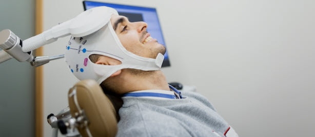
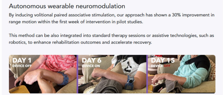
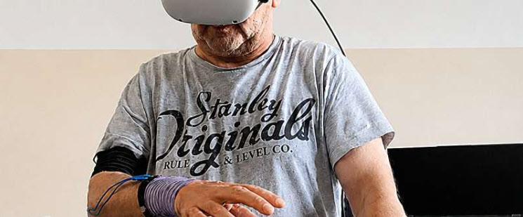
The advancement of stroke rehabilitation continues to evolve with the integration of neuromodulatory technologies designed to augment traditional physiotherapy outcomes. A prominent development in this field is the TRICEPS trial, which was recently featured on the BBC Tech Now programme, featuring Dr Sheharyar Baig, a neurologist at Sheffield Teaching Hospitals NHS Foundation Trust. This clinical investigation focuses on the efficacy of Transcutaneous Vagus Nerve Stimulation (TVNS), a non-invasive method of stimulating the vagus nerve to facilitate neuroplasticity. Unlike earlier iterations of vagus nerve stimulation that necessitated the surgical implantation of a pulse generator, the TVNS approach utilised in the TRICEPS trial employs a specialised earpiece that delivers mild electrical impulses through the skin of the ear. This stimulation is specifically timed to occur during the performance of rehabilitation exercises, with the theoretical aim of “priming” the brain to be more responsive to motor retraining.

The underlying mechanism of paired vagus nerve stimulation involves the release of neuromodulators, such as acetylcholine and noradrenaline, which are critical for strengthening the neural connections required for motor control. By delivering these pulses while a survivor engages in task-specific exercises, the technology seeks to enhance the relevance of physical therapy and improve upper limb function. Clinical accounts from the trial, such as that of participant Amanda, illustrate the practical implications of this research. After six years of persistent right-hand impairment, Amanda reported significant functional gains following a 12-week regimen, which involved using the device for approximately one hour daily. These gains included the restoration of fine motor skills required for sewing and the ability to perform activities of daily living, such as dressing and housework, without assistance.

The TRICEPS trial is currently the largest study of its kind in the UK, operating across 19 NHS hospital sites, including the Royal Hallamshire Hospital in Sheffield. Following an independent review, the recruitment target has been expanded to 270 participants to ensure the statistical robustness of the final results. Eligibility is broad, including adults who have experienced an ischemic stroke between six months and ten years ago and who still suffer from moderate to severe arm weakness. As recruitment is scheduled to conclude in July, the trial organisers are actively seeking final participants to help establish whether this cost-effective, at-home solution can be scaled nationally. Interested survivors can apply through the Trial Manager, Kirsty McKendrick, or contact the team at triceps@sheffield.ac.uk.

While the majority of stroke survivors battle the well-documented hurdles of hemiparesis or aphasia, a small fraction of individuals find themselves navigating bizarre neurological landscapes that defy conventional understanding. One of the most striking is Alien Hand Syndrome, where a limb appears to act with a mind of its own, performing complex tasks like buttoning a shirt or grabbing objects without the person’s conscious intent. This dissociation between will and action is estimated to affect fewer than one per cent of stroke patients, yet for those involved, it creates a profound sense of physical betrayal. Because the condition is so rare and often misdiagnosed, only a handful of survivors ever receive the specialised sensory-motor training required to dampen these involuntary movements.

Equally disorientating is Alice in Wonderland Syndrome, a perceptual disorder where the survivor perceives parts of their own body or external objects as being wildly out of proportion. A hand might appear as large as a car, or the floor may seem to undulate like water, creating a terrifying lack of spatial stability. Research suggests this occurs in a very low percentage of cases, primarily when the stroke affects the parietal or occipital lobes. Despite the severity of the distortion, many patients are reluctant to seek help, fearing that their symptoms will be mistaken for psychiatric illness rather than a secondary effect of neural misfiring.

In some instances, the stroke can even alter a person’s perceived identity through Foreign Accent Syndrome. This occurs when damage to the speech centres of the brain changes the rhythm, pitch, and intonation of speech, causing the survivor to speak with a distinct accent they have never naturally possessed. While it is often treated as a media curiosity, it is a genuine clinical problem that affects roughly one in a thousand survivors. The psychological impact of losing one’s native voice is immense, yet specialised speech therapy designed specifically for this syndrome is incredibly difficult to access in the UK, leading to a low rate of successful intervention.

Even more elusive are Prosopagnosia and Cotard’s Delusion, both of which strike at the heart of human recognition and existence. Prosopagnosia, or face blindness, leaves a survivor unable to recognise the faces of their own family members or even their own reflection, despite their vision remaining perfectly clear. On the extreme end of the spectrum is Cotard’s Delusion, a nihilistic belief where the patient is convinced they have died, do not exist, or have lost their internal organs. These conditions are so exceptionally rare that they appear in fewer than 0.5 per cent of clinical cases. Consequently, the vast majority of those suffering do so in isolation, as standard rehabilitation units are rarely equipped to address these profound cognitive shifts.

Ultimately, these conditions represent the fringes of neurological recovery in 2026. While the chance of encountering them is statistically low, they are a daily reality for a resilient few who must find ways to cope with a world that has become unrecognisable. Because these problems are so rare, clinical data on successful management is scarce, making it vital for survivors to share their personal strategies. Do any of our readers suffer with these unusual limitations or perhaps something similarly rare?? If you have found a way to manage these unique challenges, please let us know in the comments below how you cope so that others might benefit from your experience!


We are on Facebook

ARNI