Your Stroke / Brain Injury Recovery Starts Here


ARNI home-based training and guidance for your rehab is POWERFUL. Accept no substitute.

2026 marks 25 years of ARNI: Click Here to Claim your £50 off Full Set of Stroke Rehab 7 DVDs TODAY!

News

Researchers at Duke University School of Medicine have published findings in Nature describing a technology called LinCx that may have direct relevance to stroke survivors.The basic idea is this: if a brain connection is damaged, instead of trying to fix it or medicate around it, you build a new electrical path between the neurons that need to talk to each other. Like dealing with a road closure, LinCx creates a working detour with a biological ‘wire” designed to bypass broken or disrupted brain connections.

Stroke frequently does not kill every neuron in the affected area; what it does in many cases is sever the connections between neurons that remain alive and present. Those disconnected cells can no longer communicate reliably – and it is that loss of communication, rather than simple cell death, that also underlies persisting problems after stroke: like fatigue, low mood, cognitive difficulty etc. LinCx, led by Kafui Dzirasa at Duke, does not attempt to repair damaged synapses; instead it builds a new electrical ‘bypass’ between specific neurons, restoring signal transmission without altering the brain’s existing connections.

The system uses proteins originally identified in fish, where they function naturally as electrical synapses. The Duke team engineered these proteins to bond only with matching modified partners, preventing interaction with the brain’s natural proteins and avoiding unintended connections elsewhere. Earlier attempts to exploit electrical synapses in research settings frequently produced connections in unintended locations; LinCx addresses this through a lock and key mechanism in which each engineered protein recognises only its designated partner. Where current treatments – antiepileptic drugs, antidepressants, electrical stimulation devices – act across broad areas of neural tissue, LinCx targets selected neurons only. In animal testing, the added connections strengthened communication in specific circuits, altered brain-wide activity patterns, and produced measurable behavioural changes related to stress responses and social interaction across two different species.

The implications for stroke survivors are worth considering: post-stroke epilepsy, affecting approximately one in ten survivors, arises from disordered electrical activity in damaged circuits; current treatment suppresses symptoms pharmacologically without addressing the underlying disruption. Post-stroke depression, which affects around a third of survivors, involves significant circuit level dysfunction in the connections between the prefrontal cortex and subcortical mood regulation structures – connections that medication restores only partially. Cognitive impairment, spasticity and post-stroke fatigue similarly reflect disrupted circuit communication rather than simply lost tissue; and in each case a technology capable of restoring specific connections rather than broadly modulating the nervous system.

LinCx remains at the animal research stage. Dzirasa has indicated that the next step is establishing whether the technology can override synaptic deficits caused by lifelong genetic disruptions – still considerably short of human trials. The UK regulatory pathway through the MHRA, encompassing safety, tolerability and efficacy phases, typically requires ten to twenty years for a novel biological technology. The research is funded by the Howard Hughes Medical Institute, the National Institutes of Health, the Burroughs Wellcome Fund and the Hartwell Foundation. Routine NHS availability before the late 2030s is unlikely; the 2040s is a more grounded estimate.

The integration of autonomous gyroscopic stabilisation and artificial intelligence has established a new academic benchmark for the management of truncal ataxia following a neurological event. It’s clear that traditional walking aids (such as the heavy rollators often used by younger survivors) fail to address the underlying cerebellar dysfunction that leads to coordination failure; this limitation occurs because static frames provide a cumbersome external support rather than active stabilisation. This research into the Gyropack – conducted by Erasmus MC, Radboudumc, and TU Delft – utilised high-speed flywheels similar to those used to stabilise satellites in space… and it discovered that the recruitment of these ‘control moment gyroscopes’ allows the torso to remain upright by providing resistance to rapid, unstable movements.

In many cases; the use of a walker is experienced as stigmatising and awkward; this phenomenon suggests that a wearable solution is essential for restoring both physical freedom and social confidence. But when a device like the Gyropack (developed by Heike Vallery and Bram Sterke) uses rapidly spinning flywheels to create a sensation akin to moving in water; it takes over the heavy lifting of torso control by slowing down trunk oscillations. So you’ve got a situation where the success of your balance depends on having more time to adjust your own posture before a fall occurs.

The engineering specifically looks at how ‘active’ gyroscopic resistance (delivered via two cylinders or ‘soup cans’ containing motors and flywheels) affects your ability to walk in a straight line. You’ve got to consider that even the inactive weight of the six-kilogram pack provides some passive stability; however, principal investigator Jorik Nonnekes found that the active mode produced the greatest improvement in gait for those with moderate to advanced ataxia. And by utilising these aerospace principles; researchers can now provide a stabilising force that exists entirely within the user’s center of gravity. This points us toward a model of wearable autonomy where the backpack acts as a personal balancing aid (effectively replacing the need for a walking frame at social events).

Implementation of the Gyropack into routine NHS clinical practice in the UK remains a distant prospect; it will likely require another five to ten years to address current issues with noise and weight for a start (it currently feels like wearing a vacuum cleaner!) before it is suitable for a trip to the supermarket. But the foundational science is now solid… so the focus is now to making the system lighter and quieter for daily use. You’ve effectively seen the first steps toward a future where aerospace engineering preserves quality of life!

Recent pharmacological developments at the University of Hong Kong under the direction of Professor Aviva Chow Shing-fung have provided a sophisticated analysis of how nanotechnology can facilitate immediate neuroprotection following a stroke. So, the failure rate of drugs targeting the central nervous system exceeds 90 per cent because the blood-brain barrier acts as a formidable gatekeeper; however, the traditional view of hospital0bound treatment is being challenged by a more nuanced understanding of the nose to brain pathway.

This research apparently utilised a ‘Nano-in-Micron’ delivery system to monitor how inhalable particles can bypass systemic circulation while survivors are still in transit to the hospital… and it discovered that the recruitment of these nanoparticles allows for the direct delivery of medication to damaged cerebral tissue. In many cases, the death of brain cells occurs within minutes of an ischemic event; this phenomenon, known as the ischemic cascade, suggests that early intervention is the only way to preserve the remaining healthy tissue in your neural networks. But when the drug is delivered via a nasal spray, it travels along the olfactory and trigeminal nerves to take over the heavy lifting of cellular protection before you even reach the ward. So you’ve got a situation where the success of your rehabilitation depends entirely on whether your brain receives stabilisation in a productive or a counter-productive timeframe…

The study specifically looked at how ‘Nanopowder’ integrity, which acts like an express lane to the brain, affects your ability to maintain neurological and bodily functions. Consider, that if the intervention occurs within 30 minutes, the brain will seek to maintain its integrity and reduce tissue death by over 80 per cent. And so, by mapping these delivery pathways, researchers can now now predict how to mitigate inflammation and prevent apoptosis during the critical golden hour. This points us toward a model of prehospital precision medicine where the timing of the nasal dose dictates the exact extent of stroke survivors’ long-term recoveries.

Implementation of these specific nasal protocols into routine NHS clinical practice in the UK remains a distant prospect; it will likely require at least another five to ten years of largescale longitudinal studies to validate the cost-effectiveness of such high-level emergency tools. But the foundational science is now solid… so the focus is shifting toward developing portable rescue kits that might one day protect these specific brain regions during your journey in the ambulance.

The CHOICE2 trial coordinated by Hospital Clinic Barcelona and IDIBAPS has provided a rigorous academic basis for a shift in how we’ve traditionally viewed the limitations of mechanical thrombectomy. While the removal of a primary thrombus via a stent retriever has been the standard of care for severe ischemic stroke, this study published in JAMA confirms that simply reopening a major vessel is often insufficient for complete tissue reperfusion.

If you’ve had one of these, you also possibly may have experienced the reality where a technically successful procedure does not translate into functional independence; this discrepancy can occur for many reasons – and one of them has been found to be due to microcirculatory failure in the smaller distal vessels. The research team led by Professor Angel Chamorro and Dr Arturo Renu utilised a randomised clinical trial involving 440 patients to demonstrate that the targeted administration of intra -arterial alteplase immediately following a thrombectomy significantly improves outcomes.

This pharmacological intervention acts upon the ‘side streets’ of the cerebral vasculature… and it ensures that the microcirculation is cleared of the residual fibrin that the mechanical device cannot reach. The trial showed a marked increase in the percentage of patients achieving a mRS score of 0 to 1 at ninety days… moving from 42.5 per cent in the control group to 57.5 per cent in the treatment group. But the success of this combined strategy depends on the precision of the delivery during the angiography… so the drug is applied directly to the affected territory to avoid the systemic risks associated with traditional thrombolysis. Professor Chamorro’s findings suggest that we are entering an era of secondary reperfusion strategies where the focus moves from the macrovascular level to the cellular level of the brain tissue itself.

The data indicates that there was no significant increase in symptomatic intracranial haemorrhage; which tends to reduce the risks of combining surgery with potent lytics. In the UK, the adoption of the CHOICE2 protocol into NHS guidelines will likely require further health economic assessments to justify the additional time spent in the catheterisation suite. It is anticipated that this evidence will begin to influence British specialist stroke centres within the next three to five years as international consensus statements are updated.

Recent neuroimaging studies conducted at Syracuse University under the direction of Professor Ellyn Riley have provided a sophisticated analysis of how your brain attempts to reorganise itself following a left hemisphere stroke. So, aphasia occurs when the primary language centres are damaged; however, the traditional view of the right hemisphere simply acting as a dormant backup is being replaced by a more nuanced understanding of neural plasticity. This research apparently utilised functional magnetic resonance imaging to monitor blood flow and neural activation patterns while survivors performed linguistic tasks… and it discovered that the recruitment of the right hemisphere is not always a helpful adaptation.

In many cases, the right side of your brain might actually interfere with the recovery of the left side; this phenomenon, known as transcallosal inhibition, suggests that certain neural compensations can actually suppress the remaining healthy tissue in your speech centres. But when the damage to your left hemisphere is too extensive, the right hemisphere must step in to take over the heavy lifting of processing syntax and vocabulary. So you’ve got a situation where the success of your rehabilitation depends entirely on whether your brain is reorganising in a productive or a counter-productive manner.

The study specifically looked at how white matter integrity… which acts like the wiring between different brain regions… affects your ability to regain fluent speech. You’ve got to consider that if the structural pathways are too degraded, the brain will seek less efficient workarounds that produce slower, more effortful communication. And by mapping these pathways, researchers can now predict which types of intensive speech and language therapy will work best for your specific brain profile.

This points us toward a model of precision medicine where the location and volume of your lesion dictate the exact frequency and type of therapeutic intervention you receive. Implementation of these specific imaging protocols into routine NHS clinical practice in the UK remains a distant prospect; it will likely require at least another five to ten years of largescale longitudinal studies to validate the cost – effectiveness of such high -level diagnostic mapping. But the foundational science is now solid… so the focus is shifting toward developing wearable technologies that might one day stimulate these specific brain regions during your daily exercises. You’ve effectively become a participant in a global effort to decode the limits of human neurobiology! 

The CorTec Brain Interchange system’s neuroprosthetic is designed to function as a fully implanted wireless closed-loop platform that monitors and stimulates the brain simultaneously. You’ve probably seen various brain–computer interfaces in the news… but this specific hardware is unique because it avoids the need for external cables or bulky headgear that typically limits a survivor’s mobility during rehabilitation.

The techutilises ‘Air–Ray’ electrodes embedded within soft silicon sheets which are designed to rest upon the cortical surface rather than penetrating the brain tissue; this is a critical safety feature for long term use as it reduces the risk of inflammation or scarring whilst maintaining a high resolution for neural signal acquisition. In a notable clinical trial at the University of Washington, a participant was able to play the video game ‘Pong’ using nothing but his thoughts just two hours after the system was introduced. This speed of acquisition suggests that the decoding algorithms used to interpret your intent to move are becoming incredibly efficient. Dr Frank Desiere and his team have ensured the hardware remains identical for both the rehabilitative stimulation and the digital control aspects; so you’ve no requirement for additional surgical interventions or separate devices to switch between physical therapy and computer interaction.

The system has achieved an ‘FDA Breakthrough Device Designation’ in the United States… which accelerates the regulatory path for technologies that provide more effective treatment of life–threatening or irreversibly debilitating conditions. Prof Jeffrey Herron and other leading researchers involved in the study believe the ability to record and stimulate in real time allows for a more tailored approach to neuroplasticity. By delivering precisely timed electrical pulses… the device aims to strengthen the neural pathways you’ve lost after a stroke; essentially helping the brain to rewire itself more effectively than through traditional passive exercises alone. Although the primary trials are taking place across the Atlantic… the German engineering firm CorTec is actively looking toward the European and British markets for future routine implementation.

It’s difficult to pin down a precise date for when you might see this in a standard UK clinical setting; however the progression of clinical programmes for paralysis and the move toward more personalised neurotherapies suggest we are looking at a timeframe of several years rather than decades. The transition from feasibility studies to widespread NHS availability will depend heavily on larger scale trials proving long term efficacy and cost-effectiveness. ARNI Stroke Rehab Ul says that the dual capability of brain stimulation and digital control mean that closed–loop neurotherapies could possibly be moving closer to becoming a standard part of long term recovery in the future, but whilst the technology is promising, there’s currently no firm date for when such advanced systems will be available for routine use in the UK. Interesting however to know that this tech is out there and hopefully coming our way….

The ESPRESSo trial – Enhancing Spontaneous Recovery after Stroke – published in March 2026 in Brain Communications, delivers a clear finding: adding 90 minutes of high-intensity upper limb therapy each weekday for three weeks, begun within two weeks of stroke, did not produce better three-month outcomes than standard care alone.

Led by Professor Winston D. Byblow of the Department of Exercise Sciences and Centre for Brain Research at the University of Auckland, alongside Professor Cathy M. Stinear, Professor P. Alan Barber, and Professor John W. Krakauer of Johns Hopkins University, the trial enrolled 64 stroke survivors; half used a MindMaze videogame platform generating high volumes of exploratory hand and arm movements, half received conventional therapy… neither group outperformed a historical cohort who had received standard care only. The trial ran at Auckland City Hospital between 2021 and 2024 and was funded by the Health Research Council of New Zealand. Outcomes were measured immediately after intervention, then at three and six months post-stroke, with the primary endpoint being upper limb capacity at three months on the ARAT (Action Research Arm Test). Both intervention groups improved markedly between the start of the study and the end of the additional therapy period, with further smaller gains at three months,but those three-month outcomes were indistinguishable from the standard care cohort.

What makes this significant? The assumption that earlier and more intensive therapy capitalises on a window of neural plasticity has shaped stroke rehabilitation for years (partially dismantled by AVERT). But as Professor Byblow states: ‘early recovery after stroke is dominated by powerful biological repair processes, and increasing therapy dose very early after stroke may not enhance those processes.’ You’ve basically got a brain already doing a great deal of the work; and the evidence here suggests pushing harder against that process adds nothing at that early stage.

Crucially, participants were selected using biomarker stratification of corticospinal tract integrity (a world-first for a rehab trial), so the null result cannot be attributed to mixing high and low potential responders. It’s also worth noting that the videogame platform was rated as enjoyable by patients and achieved the same outcomes as conventional therapy; so digital rehab tools need not be inferior to hands-on treatment, even if neither added benefit over standard care at this early stage. Professor Byblow has suggested that the biggest gains from intensive training may come after spontaneous biological recovery has run its course, and that exploring biological treatments in the acute phase may be more productive than simply increasing activity-based loading.

This connects to the Auckland group’s longer body of work on proportional recovery; the finding that most survivors recover approximately 70% of lost upper limb function within three months if their corticospinal tract remains sufficiently intact. Biomarker tools such as the PREP2 algorithm – combining TMS-derived motor evoked potential data with clinical assessment – already predict upper limb recovery with over 80% accuracy and have been adopted at Auckland City Hospital.

In the UK, TMS is not yet close to being any kind of standard intervention in stroke units and NHS-wide biomarker-guided rehab remains many years off (something that ARNI friend (Emeritus) Professor Val Pomeroy is examining right now (and has done for many years). Realistically, routine adoption is unlikely before the early 2030s.

The persistent hand weakness that drives the need for trials like ESPRESSo is well documented; it’s known to reduce independence at six months after stroke and remains one of the most clinically significant unresolved challenges in neurorehabilitation. If the early acute phase is dominated by biology rather than therapy dose, the implication for NHS stroke services is that resource in the first two weeks might be better directed toward fatigue management, patient readiness, and preparing for the more intensive rehabilitation work that may yield greater returns once spontaneous recovery has stabilised.

ARNI Stroke Rehab UK says: this research reinforces that working with your biology rather than just against your deficits… and knowing where you are in your recovery trajectory… is what will make the real difference to what you get back.


Stroke recovery research has long focused on what the damaged brain can no longer do. A study published in The Lancet Digital Health by scientists at the USC Mark and Mary Stevens Neuroimaging and Informatics Institute (Stevens INI) shifts that focus considerably – finding that the undamaged side of the brain may actively reorganise itself after stroke, showing signs of ‘younger’ biological structure as it adapts to injury… and the implications for how we understand neuroplasticity and rehabilitation are meaningful.

The research is part of the ENIGMA Stroke Recovery Working Group, a global collab led by Dr. Hosung Kim, Professor of Research Neurology at the Keck School of Medicine of USC, and Dr. Sook-Lei Liew at the Stevens INI at the USC Viterbi School of Engineering. The dataset analysed brain scans from more than 500 stroke survivors across 34 research sites in eight countries, building what is described as the world’s largest stroke neuroimaging dataset of its kind. That scale matters enormously – the subtle patterns this study identified would simply not be detectable in the smaller, single-site studies that have historically dominated stroke neuroimaging research.

The analytical method at the heart of the study is worth understanding. The team used a graph convolutional network – which is basically an advanced form of AI, trained on tens of thousands of MRI scans …to estimate the biological age of 18 distinct brain regions in each hemisphere. The difference between a person’s predicted brain age and their actual chronological age is known as the brain-predicted age difference (brain-PAD), and it functions as a sensitive marker of neural health. An older-appearing brain-PAD has previously been associated with Alzheimer’s disease, traumatic brain injury and major depression, while a younger-appearing brain-PAD suggests preserved structural integrity. And, what made this study striking was what happened on the undamaged side…

In survivors with the most severe movement deficits – even after six or more months of rehabilitation – the damaged hemisphere showed accelerated ageing consistent with the scale of the lesion, as you’d expect. But the opposite hemisphere, particularly within the contralesional frontoparietal network, showed a paradoxically younger-than-expected brain age. The frontoparietal network is known to support motor planning, attention and coordination; it’s not the primary motor system but it plays a critical compensatory role when the primary motor pathways are damaged. As Dr Kim explained, rather than indicating full recovery of movement, this younger pattern likely reflects the brain’s attempt to adjust when the damaged motor system can no longer function normally… and that distinction is clinically important because it means the pattern is a marker of compensation rather than recovery, which has direct implications for how rehabilitation targets should be set.

Dr Liew noted a paradoxical finding; that the contralesional rejuvenation effect was strongest in the most severely impaired survivors, suggesting that the greater the demand placed on the intact hemisphere by severe ipsilesional damage, the more structurally ‘youthful’ those compensatory regions appear. This is btw, consistent with use-dependent plasticity (where regions under higher functional demand maintain or enhance their structural integrity). Professor Arthur W. Toga PhD, director of the Stevens INI, added that pooling worldwide data and applying AI allows detection of patterns invisible in smaller studies, and that these findings could eventually guide personalised rehabilitation strategies. For now brain-PAD remains a research tool, and translating it into routine NHS practice requires replication and NICE review – a process measured in years rather than months. But the message is clear; the brain after stroke is not passively awaiting recovery.. it’s actively reorganising – and understanding that process more precisely is likely to be one of the threads in stroke rehab research over the next decade.

Chronic pain is one of the most prevalent and least adequately treated sequelae of stroke. Research suggests that approximately 42% of stroke survivors are living with chronic pain six months or more after their stroke… and with around one million stroke survivors in the UK, that points to roughly 420,000 people managing pain as a direct or indirect consequence of their stroke, a number projected to grow substantially as the stroke survivor population is expected to more than double by 2035.

The pain broadly divides into two categories. Central post-stroke pain (CPSP) arises directly from damage to the spinothalamic pathways and thalamic structures involved in pain processing; it’s characterised by burning, aching or hypersensitivity and responds poorly to conventional analgesics. Musculoskeletal pain- including chronic low back pain – arises from the biomechanical consequences of stroke; altered gait, spasticity, pelvic tilt, inhibited core and gluteal muscles, and hours spent in poorly supported positions all drive cumulative mechanical stress into the lower spine over months and years. Lower back pain in particular becomes a significant independent barrier to rehabilitation, reducing exercise tolerance, disrupting sleep and compounding fatigue.

Current treatment options are limited. Opioids carry serious risks of dependence and cognitive blunting, especially concerning in a population already managing potential cognitive impairment. Existing implantable spinal cord stimulators work but require invasive surgery, battery replacement and are expensive… none of which makes them easily accessible to most stroke survivors.

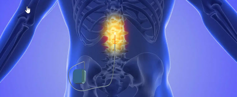
This is why the work from Professor Qifa Zhou at the Zhou Lab, USC Viterbi School of Engineering, published in Nature Electronics in June 2025 and developed in collaboration with the Jun Chen Group at UCLA – deserves attention. Lead author PhD candidate Yushun Zeng and the team have developed a flexible ultrasound-induced wireless implantable (UIWI) stimulator that receives power entirely from a wearable external ultrasound transmitter worn over the skin, with no batteries, no wires and no repeated surgery. The device uses the piezoelectric effect – converting ultrasound mechanical waves into electrical stimulation delivered to the spinal cord – and integrates machine learning algorithms that read physiological signals in real time and adapt stimulation continuously rather than delivering a fixed response. For a stroke survivor whose pain fluctuates with activity, fatigue and spasticity throughout the day, that adaptive capacity is definitely a serious clinical step beyond anything currently available.

Routine clinical use in the UK will be some years away; MHRA licensing and NICE appraisal following human trials would realistically place this five to eight years from now at minimum. But the direction is clear, and that matters. ARNI Stroke Rehab & Recovery says: chronic lower back pain is one of the most consistently reported barriers to rehabilitation that stroke survivors describe to us, and drug-free adaptive pain technology like this represents exactly the kind of progress that could help people keep moving, keep training and keep improving long after their stroke.

Measuring stroke recovery properly is harder than it sounds. Most single outcome measures capture one dimension of what stroke does to a person and miss everything else… which is why Dr Tom Balchin at ARNI Stroke Rehab UK has built a set of four complementary assessments into every ARNI specialist’s training and into the Training Logbooks that survivors and instructors use together, repeated every 12 weeks so that genuine progress – and any regression – can be tracked with rigour over time.

The Stroke Impact Scale (SIS) is introduced from day one of the ARNI Functional Rehabilitation after Stroke Accreditation; it covers strength, memory, emotion, communication, activities of daily living, mobility and quality of life, giving both trainer and survivor a rounded picture of how stroke is affecting daily living across every domain. Alongside it, also from the outset, comes the Chedoke Arm and Hand Activity Inventory (CAHAI); it assesses the functional use of the affected arm and hand across nine real-life bilateral tasks — opening a jar, doing up buttons, pouring a glass of water and more – producing a score out of 63 that reflects what the upper limb can actually do in practice rather than just what it looks like in a clinical setting. Together the SIS and CAHAI form a strong and complementary pairing; the SIS capturing the broad whole-life picture and the CAHAI drilling down into the upper limb detail that sits at the heart of so much of ARNI’s rehabilitation work.

Once students are established in the course, the Rivermead Mobility Scale (RMS) is introduced; it measures mobility across 15 tasks from turning over in bed through to running, making it an ideal tool for tracking lower limb and whole body functional progress over time. And most recently Dr Tom has begun introducing instructors to the Fatigue Severity Scale (FSS), developed by Krupp et al… which was advised to him by his kind contact Dr Anna Kuppuswamy (formerly of Queen Square, UCL and now at Leeds), because post-stroke fatigue is one of the most debilitating and underassessed sequelae of stroke, recognised by survivors themselves as something that can affect them every day.

What seems to make this quad quite effective is that together they cover the full landscape of stroke recovery without much overlap – upper limb function, mobility, whole-life impact and fatigue – and no single measure could do that alone. They are the evidence base that shows survivors, families and commissioners what ARNI training is actually achieving, session by session and month by month. Every ARNI instructor is trained by Dr Tom and his team to use them with care and consistency.


We are on Facebook

ARNI